In the ever-evolving field of dentistry, technological advancements play a crucial role in enhancing patient care and treatment outcomes. One such significant innovation is digital x-ray technology, which has revolutionized the way dental professionals diagnose and treat various oral health issues. Unlike traditional film x-rays, digital x-rays offer numerous benefits, including enhanced image quality, reduced radiation exposure, and improved diagnostic capabilities. In this blog, we will explore the latest advancements in digital x-ray technology and discuss how they are transforming dental practices and improving patient experiences.
In This Blog:
- What is Digital X-Ray Technology?
- Advantages of Digital X-Ray Technology
- Technological Advancements in Digital X-Rays
- Impact on Dental Practices
What is Digital X-Ray Technology?
Digital x-ray technology represents a significant advancement over traditional film-based x-rays, providing a host of benefits for both dental professionals and patients. Unlike conventional x-rays, which use film to capture images, digital x-rays utilize digital sensors or phosphor plates to create images that are immediately available for viewing on a computer screen. This section will explore the fundamental aspects of digital x-ray technology, how it works, and the various types of digital x-ray systems available.
Explanation of Digital X-Rays Compared to Traditional Film X-Rays
Traditional film x-rays have been a cornerstone of dental diagnostics for many decades. They involve exposing a piece of photographic film to x-ray radiation, which then needs to be developed using chemical processes. This method, while effective, has several drawbacks, including higher radiation exposure, longer processing times, and the need for physical storage of x-ray films.
Digital x-rays, on the other hand, employ electronic sensors or phosphor plates to capture images. When the x-ray beam passes through the patient’s body, it is detected by the sensor or plate, which then converts the data into a digital image. This image can be viewed almost instantly on a computer monitor, allowing for quicker diagnosis and treatment planning.
How Digital X-Rays Work
Digital x-rays function by utilizing either direct or indirect methods to capture images. In the direct method, a digital sensor is placed inside the patient’s mouth to capture the x-ray image directly. This sensor is connected to a computer, which immediately displays the image. In the indirect method, a phosphor plate is used to capture the image, which is then scanned into a digital format using a special scanner.
The process begins with the patient being positioned in the x-ray machine, just as with traditional x-rays. However, instead of using film, the x-ray beam strikes the digital sensor or phosphor plate. These devices are sensitive to x-rays and convert the radiation into electrical signals, which are then processed by software to create a detailed digital image. These images can be enhanced, enlarged, and manipulated to aid in diagnosis.
Types of Digital X-Ray Systems
There are several types ofdigital x-ray systems used in dentistry, each with its specific applications and benefits:
Intraoral X-Rays:
These are the most common type of dental x-rays, used to capture images of individual teeth and surrounding bone structures. Intraoral x-rays provide detailed images that are essential for diagnosing cavities, tooth infections, and other dental issues.
Extraoral X-Rays:
These x-rays capture images of the entire jaw and skull. They are typically used to examine the larger structures of the mouth and face, such as the jawbone and temporomandibular joints (TMJ).
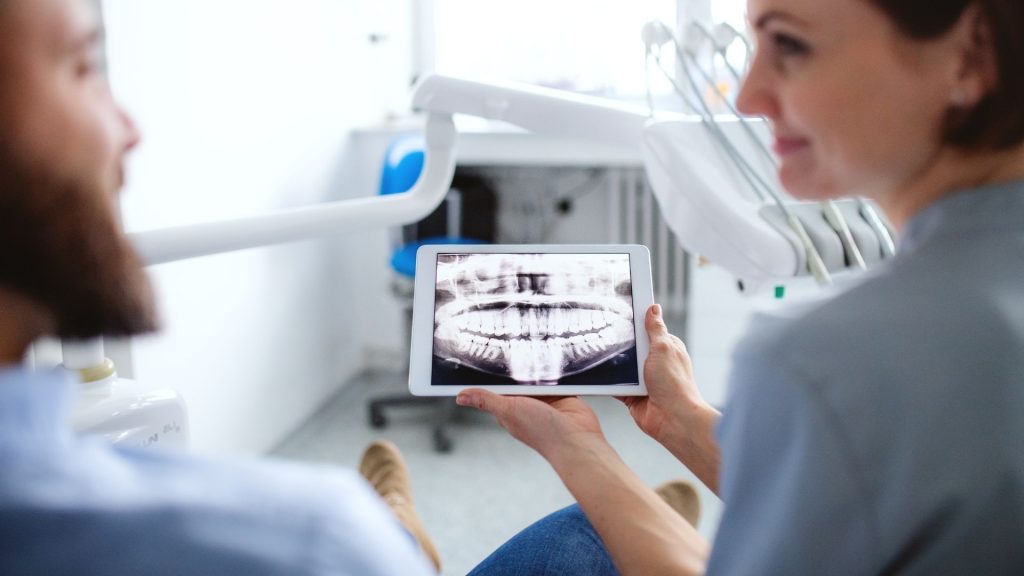
Panoramic X-Rays:
Panoramic x-rays produce a single image of the entire mouth, including the teeth, jaws, and surrounding structures. This type of x-ray is particularly useful for planning dental implants, detecting impacted teeth, and assessing jaw alignment.
Cephalometric X-Rays:
These specialized x-rays capture side views of the head and are commonly used in orthodontics to evaluate jaw relationships and plan orthodontic treatments.
By understanding the different types of digital x-ray systems and how they function, dental professionals can choose the most appropriate technology for their diagnostic needs, ultimately improving patient care and treatment outcomes.
Advantages of Digital X-Ray Technology
The shift from traditional film-based x-rays to digital x-ray technology has brought about a multitude of benefits that enhance both the quality of dental care and the overall patient experience. This section will delve into the key advantages of digital x-ray technology, highlighting its superiority in terms of image quality, safety, efficiency, and environmental impact.
Enhanced Image Quality
One of the most significant advantages of digital x-rays is the superior image quality they provide. Digital x-rays offer higher resolution images, allowing for greater detail and clarity. This enhanced image quality improves diagnostic accuracy, enabling dental professionals to detect issues that might be missed with traditional film x-rays. Furthermore, digital images can be easily adjusted for brightness, contrast, and zoomed in for closer inspection, facilitating better analysis and more precise treatment planning.
Reduced Radiation Exposure
Safety is a paramount concern in any medical setting, and digital x-rays significantly reduce the amount of radiation exposure to patients compared to traditional film x-rays. Studies have shown that digital x-rays can reduce radiation exposure by up to 80-90%, making them a safer option for both patients and dental staff. This reduction in radiation is particularly beneficial for patients who require frequent x-rays, such as those undergoing orthodontic treatment or monitoring the progress of dental conditions.
Speed and Efficiency
Digital x-rays streamline the imaging process, making it much faster and more efficient than traditional methods. With digital systems, images are captured and displayed on a computer screen almost instantly, eliminating the need for time-consuming film development. This immediate availability of images allows dental professionals to quickly diagnose issues and discuss treatment options with patients during the same visit, reducing wait times and improving the overall patient experience. Additionally, the ease of storing and retrieving digital images enhances workflow efficiency within dental practices.
Environmental Benefits
The environmental impact of dental practices is an important consideration in today’s eco-conscious world. Digital x-ray technology contributes to environmental sustainability by significantly reducing the use of chemicals and materials associated with traditional film x-rays. Traditional x-ray processing requires chemical developers and fixers, which can be hazardous to the environment if not disposed of properly. Digital x-rays eliminate the need for these chemicals, resulting in a cleaner and greener dental practice. Furthermore, digital images can be stored electronically, reducing the need for physical storage space and minimizing paper waste.
In summary, the advantages of digital x-ray technology extend far beyond improved image quality. By reducing radiation exposure, enhancing speed and efficiency, and offering environmental benefits, digital x-rays represent a significant advancement in dental imaging. These benefits collectively contribute to better patient care, more accurate diagnoses, and a more efficient and sustainable dental practice.
Technological Advancements in Digital X-Rays

The realm of digital x-ray technology is continuously evolving, with numerous advancements enhancing its capabilities and applications in dental care. These innovations not only improve the accuracy and efficiency of diagnostic procedures but also expand the possibilities for treatment planning and patient management. This section will explore some of the most notable technological advancements in digital x-ray technology, including 3D imaging, improved sensors, cloud-based systems, and the integration of artificial intelligence.
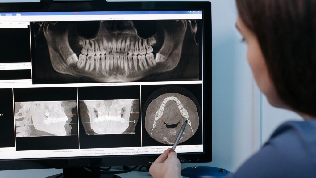
3D Imaging and Cone Beam Computed Tomography (CBCT)
One of the most groundbreaking advancements in digital x-ray technology is the development of 3D imaging and Cone Beam Computed Tomography (CBCT). Unlike traditional 2D x-rays, CBCT provides detailed three-dimensional images of the teeth, jawbone, and surrounding structures. This technology allows for a more comprehensive view of the patient’s oral anatomy, which is particularly beneficial for complex procedures such as dental implants, orthodontics, and oral surgery. CBCT can reveal anatomical details that are not visible on conventional x-rays, aiding in accurate diagnosis and precise treatment planning.
Digital Sensors and Phosphor Plate Systems
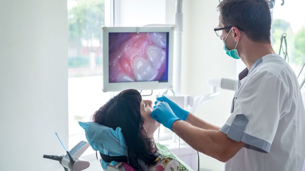
The introduction of advanced digital sensors and phosphor plate systems has significantly improved the comfort and convenience of dental x-rays for patients. Modern digital sensors are designed to be smaller and more flexible, making them easier to place inside the patient’s mouth and reducing discomfort. These sensors also provide high-resolution images with minimal radiation exposure. Phosphor plate systems, which use reusable plates to capture images, offer another option for digital imaging. These plates are scanned into a digital format, combining the benefits of digital technology with the familiarity of traditional film techniques.
Cloud-Based Imaging and Data Storage
Cloud-based imaging systems represent another significant advancement in digital x-ray technology. These systems allow dental practices to securely store and access patient images and records online, facilitating easier sharing and collaboration among dental professionals. Cloud-based storage ensures that patient data is safely backed up and can be accessed from any location, providing flexibility and convenience for both dentists and patients. This technology also supports tele-dentistry, enabling remote consultations and second opinions without the need for physical transfer of x-ray films.
Artificial Intelligence and Image Analysis
Artificial Intelligence (AI) is making its mark in the field of dental imaging, offering automated diagnostic tools and advanced image analysis capabilities. AI algorithms can analyze digital x-rays to detect early signs of dental issues such as cavities, periodontal disease, and oral cancer with remarkable accuracy. These tools assist dental professionals in making quicker and more precise diagnoses, potentially identifying problems that might be overlooked by the human eye. AI-driven image analysis also supports personalized treatment planning, ensuring that each patient receives the most appropriate care based on their unique oral health profile.
In conclusion, the technological advancements in digital x-ray technology are transforming the landscape of dental care. From 3D imaging and CBCT to improved sensors, cloud-based systems, and AI-driven analysis, these innovations enhance diagnostic accuracy, improve patient comfort, and streamline dental practice operations. As these technologies continue to evolve, they promise to further elevate the standard of dental care, making it more efficient, effective, and patient-centered.
Impact on Dental Practices
The integration of digital x-ray technology has far-reaching effects on dental practices, significantly enhancing various aspects of patient care and operational efficiency. This section will discuss how digital x-ray technology improves the patient experience, enhances diagnostic and treatment planning capabilities, and leads to cost and efficiency savings for dental practices.
Improved Patient Experience
Digital x-ray technology greatly enhances the overall patient experience in several ways. Firstly, the reduced radiation exposure associated with digital x-rays offers a safer option for patients, alleviating concerns about repeated x-ray procedures. Additionally, the comfort of modern digital sensors, which are smaller and more flexible than traditional film holders, helps to reduce patient discomfort during x-ray procedures.
The speed and efficiency of digital x-rays also contribute to a better patient experience. With images available almost instantly, patients no longer need to wait for film development, allowing for immediate discussion of diagnostic results and treatment plans. This quick turnaround not only saves time but also helps to reduce anxiety and enhance patient satisfaction. Furthermore, digital x-rays can be easily shared with patients, enabling them to better understand their oral health and treatment options through clear, high-quality images.
Enhanced Diagnostic and Treatment Planning
The high-resolution images produced by digital x-rays provide dental professionals with greater detail and clarity, leading to more accurate diagnoses. Enhanced image quality allows for the early detection of dental issues such as cavities, infections, and bone loss, which might be missed with traditional film x-rays. This improved diagnostic capability supports the development of more precise and effective treatment plans, tailored to the specific needs of each patient.
Advanced digital x-ray systems, such as Cone Beam Computed Tomography (CBCT), further enhance diagnostic and treatment planning capabilities by providing three-dimensional images of the teeth and jaw. This level of detail is invaluable for complex procedures like dental implants, orthodontics, and oral surgery, enabling dental professionals to plan and execute treatments with greater precision and confidence.
Cost and Efficiency Savings
Digital x-ray technology offers significant cost and efficiency savings for dental practices. The immediate availability of digital images eliminates the need for film development and reduces the time required for processing and handling x-ray films. This streamlining of the imaging process allows dental professionals to see more patients and spend less time on administrative tasks.
The transition to digital x-rays also reduces the costs associated with traditional film x-rays, such as the purchase of film, chemicals, and storage solutions. Over time, these savings can be substantial, making digital x-ray technology a cost-effective investment for dental practices. Additionally, the durability and longevity of digital sensors and plates reduce the frequency and cost of replacements.
Environmental benefits also contribute to cost savings. By eliminating the need for chemical developers and fixers, digital x-ray technology reduces hazardous waste disposal costs. The reduced need for physical storage space for x-ray films further contributes to operational savings.
In summary, the impact of digital x-ray technology on dental practices is profound. It improves the patient experience through enhanced safety and comfort, provides superior diagnostic and treatment planning capabilities, and offers significant cost and efficiency savings. These benefits collectively enhance the quality of dental care and streamline practice operations, positioning dental practices to deliver better outcomes for their patients.
Conclusion
The advent of digital x-ray technology has ushered in a new era of precision, efficiency, and safety in dental care. By providing high-resolution images with significantly reduced radiation exposure, digital x-rays enhance diagnostic accuracy and patient comfort. The rapid image processing and cloud-based storage options streamline practice operations, leading to cost and efficiency savings.
Furthermore, advanced technologies like 3D imaging and artificial intelligence further elevate the capabilities of dental diagnostics and treatment planning. As dental practices continue to adopt and integrate these advancements, they are well-positioned to offer superior patient care and achieve better clinical outcomes. Embracing digital x-ray technology is not just a step forward for dental practices, but a leap towards a future where dental care is more effective, patient-centered, and environmentally responsible. Come see what’s new in dental technology at Chestnut Dental!